ITB Engineering Physics Lecturers Create Imaging Devices for Medical Applications
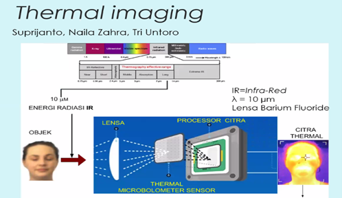
BANDUNG, itb.ac.id - Lecturers at Engineering Physics ITB have made medical imaging devices, namely "Thermal Imaging". This research was conducted by Dr. Suprijanto, S.T, M.T., Naila Zahra, S.T, M.T., and Tri Untoro, S.T, M.T.
In this device, there is a component called a thermal microbolometer. "Thermal microbolometer detects infrared radiation in our body to analyze its image," said one of ITB Engineering Physics lecturers, Dr. Vebi Nadhira, S.T, M.T., in the webinar "The Benefits of Imaging Devices in the World of Health", Tuesday (25/5/2021).
In addition to thermal imaging, the lecturers also carried out research related to medical applications, namely optical tomography, detecting discolouration of teeth, testing the spread of fluids in masks, detecting skin moisture, early detection of jaundice in infants, and needle position mapping on ultrasound images.
One of the research results on imaging in health for screening is the mapping of the needle position on the ultrasound image. This discovery was carried out by several ITB Engineering Physics lecturers and students. "This research is motivated by the implementation of ultrasound which often uses a needle to perform imaging. However, when ultrasound is performed, the inserted needle is often invisible. This research was conducted to find a technology that can make the needle on ultrasound can be seen clearly, "said Vebi. In addition, one of the mathematical formulas used in this study is a linear derivative. Linear derivatives serve to estimate or determine the area where the needle is inserted when performing an ultrasound.
Meanwhile, another research product on imaging in health for disease identification or early diagnosis is the early detection of jaundice in infants. The method used in this study is called the Kramer Method. "Before going to the doctor, the Kramer method can do imaging to detect jaundice in babies," stated Vebi.
The sample data for this study were obtained by taking ten images from 49 infant patients. To take the necessary images, a camera is placed 50 - 60 cm above the baby and a colour card at a 5 - 10 cm distance next to the baby's stomach. After getting the raw image, the filtering process is carried out and then colour correction. After that, skin segmentation was also carried out through conversion to the laboratory colour space, then switched to conversion to a specific area, acquiring statistical data on sample skin colour, and finally estimating using linear regression.
Reporter: Yoel Enrico Meiliano (TPB FTI 2020)
Translator: Billy Akbar Prabowo (Metallurgical Engineering 2020)

scan for download